Software
Description of Invention
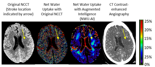
The inventors in this technology have developed software to allow the direct visualization of net water uptake in edema. In acute ischemic stroke, vessel occlusion leads to progressive edema in the brain, which is an increase of water content in the affected brain tissue. If left untreated, the progressive edema eventually becomes life threatening when tissue water content exceeds the thresholds that the specific brain tissues can hold, and the damage becomes irreversible. Therefore, water content is an important indicator of the edema level in brain tissue. Currently, net water uptake can be calculated only from a suspected stroke region that is either defined anatomically without knowledge of the stroke location or defined by other advanced imaging like CT perfusion or MRI. A brain map of net water uptake that allows direct assessment of ischemia location and extent is currently not available.
This technology combines non-contrast computed tomography (NCCT) with AI to detect the directly detect the location, extent, and level of ischemia in the brain tissue through net water uptake with augmented intelligence (NWU-AI). This type of assessment allows more sensitive and specific diagnosis of acute ischemic stroke by denoising the NCCT image by AI and the result will go through a specific process to generate a whole brain net water uptake image. In addition, NWU-AI can provide crucial information on how much tissue is salvageable or irreversibly damaged based on the level of ischemia, and it can be used to guide treatment planning for the patient even he/she is outside the traditional treatment window (e.g., 4.5 hours after stroke onset).
Stage of Development
Proof of concept retrospective studies in humans are in progress and additional prospective trials are planned.
Competitive Landscape
The current state of the field is that only specific areas of the brain are chosen, and a histogram is generated from this selected area generating only a histogram number. In contrast, this software generates a visual image of the entire scan allowing quick and easy identification of the areas of interest (the output of the process).
Intellectual Property (OTT202216)
A provisional patent application has been filed.
Inventors
Contact Us
For more information, contact the Office of Technology Transfer at OTT@HoustonMethodist.org.